Misophonia is an affective sound-processing disorder characterized by the experience of a strong negative emotional response to specific sounds, most commonly those generated when eating, drinking, and breathing; these sounds are often labeled “trigger” sounds.
The literature on misophonia has in general been limited to case studies and examination of characteristics in clinical populations, with a limited assessment of underlying physiological mechanisms contributing to the experience (Spankovich and Hall, 2014). Recently, a group out of Newcastle University in the United Kingdom, led by Sukhbinder Kumar performed functional imaging in participants with misophonia compared to matched controls.
During the imaging, participants listened to sounds rated neutral, unpleasant (but not triggers), and trigger sounds previously identified in an earlier portion of the study. Analysis of the imaging revealed significant activation of the anterior insular cortex (AIC) in response to triggering sounds compared to controls, this activation did not occur with unpleasant or neutral sounds. The AIC is involved in subjective feelings associated with emotion and a key region for detection, attention, and salience. Further, greater functional connectivity was observed in brain regions including the ventromedial prefrontal cortex (vmPFC), posteromedial cortex (PMC), and limbic area. Structural maps showed evidence of greater myelination in the gray matter of the vmPFC, supporting altered functional connectivity in persons with misophonia. Finally, physiological responses (galvanic skin response and heart rate) were correlated with activity in the AIC, which may explain anxiety or anger experienced with trigger sounds.
Overall, these new findings demonstrate a physiological basis underlying misophonia. However, these data do not differentiate the physiological and connectivity changes as a cause or a consequence of misophonia. Also, the study does not detail any overt structural abnormalities in individuals with misophonia. The question remains to what underlies this enhanced connectivity? Is this simply physiological evidence of a conditioned response? Is there some unknown pre-existing susceptibility factor to forming this connectivity (perhaps hormonal or genetic)? Can we see changes in activity after potential therapies? Hopefully, these findings will encourage support for a further study to answer these questions.
References
Kumar et al. (2017) The brain basis for misophonia, Current Biology. Online pre-publication.
Spankovich C, Hall JW. (2014) The misunderstood misophonia. Audiology Today 26(4):15–23.
Related Posts
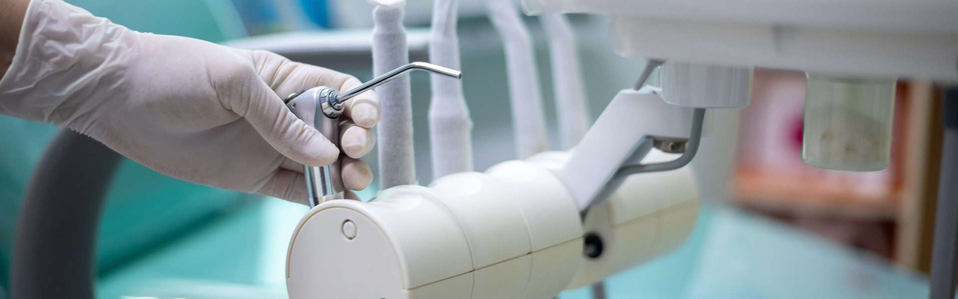
Echoes of Risk: Noise-Induced Hearing Loss in Dentistry
For audiologists, it is no surprise that dental professionals remain at risk of developing noise-induced hearing loss (NIHL). This risk is due to prolonged and…
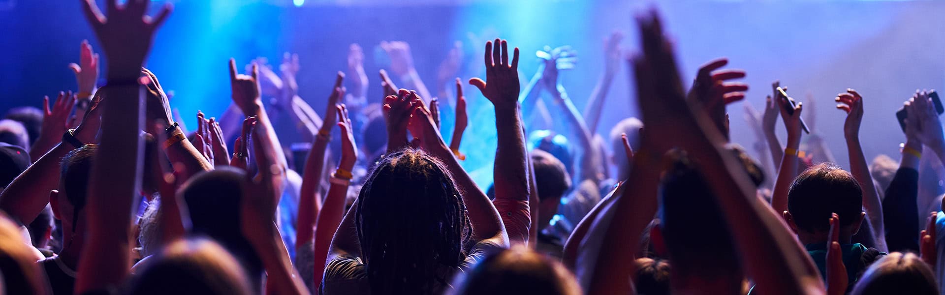
The Hobbies Most Likely to Cause Hearing Loss
Hobbies are important. They contribute to our overall health and well-being by helping us relax and escape everyday stressors. Audiologists know that some hobbies may…
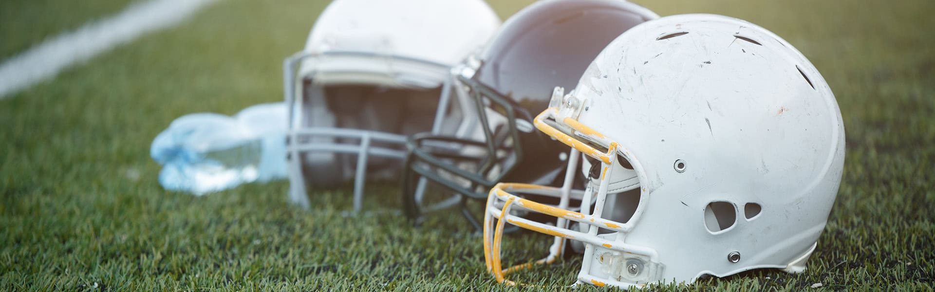
NCAA Approves Gallaudet’s Use of a Helmet for Deaf or Hard of Hearing Players This Season
A helmet designed by Gallaudet University and AT&T has been approved for use by the NCAA. Approval for the helmet in Division III college football…